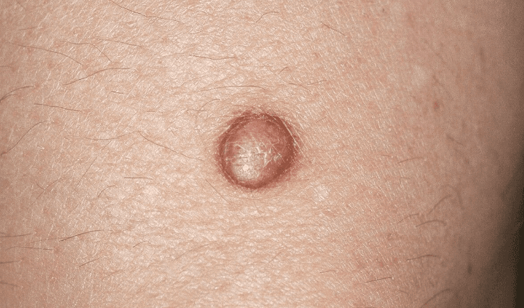
What is a dermatofibroma?
A dermatofibroma is a common, harmless skin growth: a small, firm nodule made of fibrous tissue in the dermis, the deeper of the two main layers of the skin (DermNet, BAD). It is also called a cutaneous fibrous histiocytoma or simply a histiocytoma (DermNet). Most sit on the lower legs and feel like a small rubbery button just under the skin surface (BAD). About 3 in every 100 skin lesions is a dermatofibroma (Cleveland Clinic).
Two facts matter most to patients. First, a dermatofibroma is benign and will not turn into a cancer (BAD, DermNet). Second, it can look like growths that do matter, including melanoma, so a new firm lump should still be diagnosed by a doctor rather than assumed to be harmless (BAD).
Who gets dermatofibromas?
- Adults, most often between 20 and 49. In a series of 239 dermatofibromas the average age was 41, with a range of 12 to 72 (PMC 2025 series; Cleveland Clinic).
- Women more than men. About twice as many women as men develop them (Cleveland Clinic); the 239-case series was 61% female (PMC 2025 series).
- Legs first, then arms. Half of lesions sit on the lower limbs and a third on the upper limbs; the trunk and face are uncommon sites (PMC 2025 series). In women the upper arms are a typical second site (BAD).
- All skin tones. People of every ethnicity develop dermatofibromas; the colour differs, not the growth (DermNet).
- Usually just one. Most people have a single lesion (BAD). Several appearing within a few months is unusual and is discussed below.
What causes a dermatofibroma?
The exact cause is not settled. Dermatologists still debate whether a dermatofibroma is a reaction to a small injury or a true, very slow-growing tumour of fibroblast cells (DermNet, PMC 2025 review). What is known:
- Minor skin trauma often comes first. Many appear after an insect bite, a thorn prick or an injection, although the link is not consistent (DermNet, BAD). About 1 in 5 people recall an injury at the site (Cleveland Clinic). A ruptured hair follicle is another proposed trigger (PMC 2025 review).
- It is not inherited. There is no evidence that dermatofibromas run in families (BAD).
- It is not an infection. It is an overgrowth of your own fibrous tissue (BAD), so there is nothing to pass on to anyone else.
- Many at once can point to the immune system. Many lesions (more than 15) erupting within a few months is rare and usually happens with altered immunity: HIV, immune-suppressing treatment, lupus or other autoimmune disease, and rarely certain cancers (DermNet, BAD). A doctor should look for the reason.
What does a dermatofibroma look and feel like?
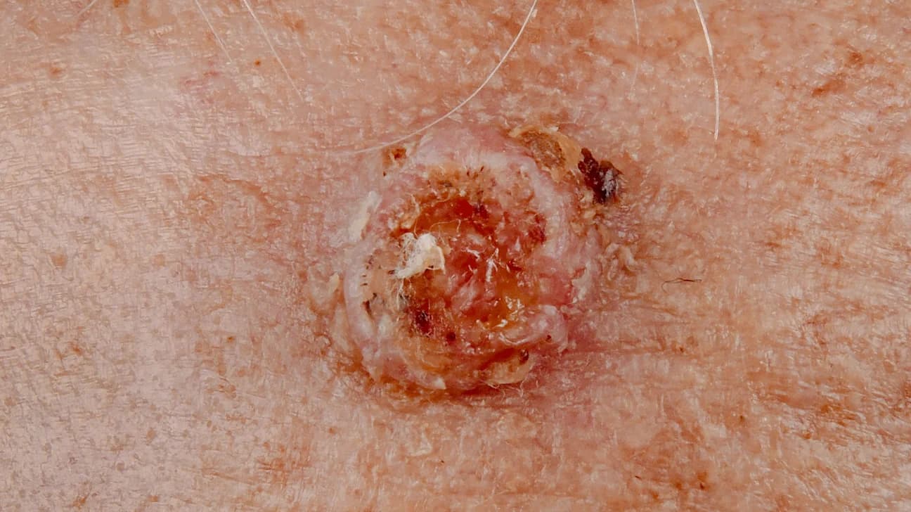
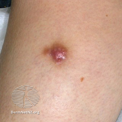
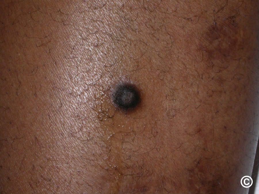
- A firm, button-like bump, slightly raised or almost flat, with a smooth surface. It is fixed to the skin above it, so the skin moves with it (DermNet, BAD).
- Small. Most measure 7 to 10 mm, within a usual range of 0.5 to 1.5 cm; most stay under 1 cm (DermNet, BAD). The average size in the 239-case series was 7 mm (PMC 2025 series).
- Colour depends on your skin. Pink, purple, light brown or grey on lighter skin; dark brown to black on darker skin. Some have a paler centre (DermNet, BAD).
- Usually no symptoms. Some people notice itching, tenderness when the lump is pressed or knocked, or occasional pain (DermNet, BAD, PMC 2025 review).
- It bleeds if a razor catches it. Because the lump stands proud of the skin, shaving over it can nick it and cause bleeding (BAD, DermNet).
- It stays put. Most dermatofibromas remain the same size for years. Growth, a change in colour or a surface that breaks down are not typical and call for a check (DermNet, PMC 2025 review).
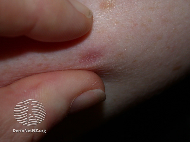
The dimple sign (pinch test)
Gently squeeze the skin on either side of the bump between your thumb and index finger. A dermatofibroma usually sinks inward and forms a dimple, because its fibrous tissue is anchored to the tissue beneath it (Cleveland Clinic, DermNet). A mole does the opposite: it stays raised, because it is not attached to the layer beneath (Cleveland Clinic).
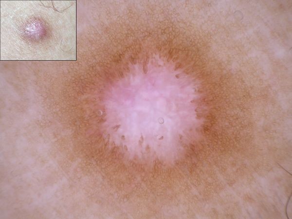
The dimple sign is a clue rather than a diagnosis. A dermatologist supports the diagnosis by examining the lesion with dermoscopy, and settles any doubt with a biopsy (DermNet).
Is a dermatofibroma dangerous? Can it be cancer?
No. A dermatofibroma is benign, does not give rise to cancer and does not spread (DermNet, BAD). The real risk is a mistaken diagnosis: a cancer that is assumed to be a harmless fibroma and left alone. Three look-alikes matter (DermNet; DermNet BCC and SCC):
- Dermatofibrosarcoma protuberans (DFSP), a rare cancer of the dermis. It affects fewer than 1 person in 100,000 a year, most often on the trunk, as a painless, rubbery, skin-coloured or red-brown plaque or nodule that keeps growing slowly over months to years and can reach several centimetres (DermNet DFSP). A fibroma that keeps enlarging needs a biopsy that reaches the deep dermis (DermNet DFSP).
- Melanoma, especially the nodular, non-pigmented (amelanotic) and desmoplastic forms, which can be pink or skin-coloured and firm (DermNet melanoma). For raised lesions dermatologists use the EFG rule: Elevated, Firm, Growing. Any lesion that changes in size, shape, colour or height for more than a month should be reviewed by a dermatologist or biopsied (DermNet melanoma).
- Basal cell carcinoma and squamous cell carcinoma, which appear on sun-exposed skin as a pearly nodule with surface blood vessels or as a tender, crusting lump that grows over weeks to months (DermNet BCC, DermNet SCC).
In doubtful cases the growth is removed and examined under the microscope. That settles the question and removes the lesion at the same time (BAD, DermNet).
Dermatofibroma or something else? A look-alike guide
A firm bump on a leg or arm has several possible explanations. A dermatofibroma is a firm, button-like nodule usually under 1 cm, fixed to the skin surface, that dimples when pinched and stays the same for years (DermNet, BAD). The table lists what sets the others apart; use it to prepare for the conversation with your doctor, not to diagnose yourself.
| Condition | How it differs from a dermatofibroma |
|---|---|
| Melanoma (nodular or amelanotic) | Elevated, firm and growing; may be pink or skin-coloured; changes over weeks to months; may bleed or crust (DermNet melanoma, NHS). |
| Dermatofibrosarcoma protuberans | Keeps enlarging, often larger than 1 cm, rubbery plaque that feels fixed; most often on the trunk (DermNet DFSP). |
| Basal cell carcinoma | Shiny or pearly nodule with fine blood vessels, often a central dip or sore that does not heal; sun-exposed skin (DermNet BCC). |
| Epidermoid cyst | Rounder and usually larger (1 to 3 cm), often with a central pore from which cheesy, foul-smelling material can be squeezed (DermNet cyst). |
| Lipoma | Soft, rubbery, lobulated lump under normal skin, often several centimetres; the skin is not tethered to it (DermNet lipoma). |
| Keloid or hypertrophic scar | Grows at the site of a cut, burn or acne spot; raised, hard, smooth and shiny; a keloid grows beyond the original wound (NHS keloid). |
| Mole, including blue naevus | Stays raised when pinched; a blue naevus is a small, smooth, bluish bump of 0.5 to 1 cm (Cleveland Clinic, DermNet blue naevus). |
| Seborrhoeic keratosis | Warty, waxy, "stuck-on" surface rather than a smooth firm nodule; very common after 60 (DermNet SK). |
| Pyogenic granuloma | Red, fleshy and fragile; grows fast over a few weeks and bleeds with minor knocks (DermNet pyogenic granuloma). |
| Prurigo nodularis | Several very itchy, scratched, firm lumps in a symmetrical pattern; driven by scratching (DermNet prurigo). |
| Persistent insect-bite reaction | Follows a bite and itches; in children bites often come in symmetrical itchy crops (papular urticaria), and a solitary reaction can persist, so the bite history matters (DermNet bites). |
| Neurofibroma | Soft rather than firm; the fingertip sinks into it through a "buttonhole" (DermNet neurofibromatosis). |
| Skin tag (soft fibroma) | Soft, skin-coloured and hanging off the skin on a stalk, usually in folds (NHS skin tags). |
Types of dermatofibroma
About 85% of dermatofibromas are the ordinary kind described above (PMC 2025 series). Pathologists recognise several variants, and a few are worth knowing because they look or behave differently (PMC 2025 review):
- Cellular dermatofibroma: more likely to reach the deepest layer of the skin and more likely to grow back after removal; still non-cancerous (Cleveland Clinic).
- Atrophic dermatofibroma: a depressed patch with a central dip rather than a bump; about 7.5% of lesions and five times more common in women (PMC 2025 review, PMC 2025 series).
- Haemosiderotic variant: a homogeneous blue, blue-red or blue-grey nodule that is often first suspected to be melanoma; the microscope gives the answer. The related aneurysmal variant contains blood-filled spaces (PMC 2025 review).
- Multiple eruptive dermatofibromas: a cluster of more than 15 lesions developing rapidly, fewer than 0.3% of cases, almost always in people with suppressed immunity (PMC 2025 review).
When to see a doctor
A stable, pea-sized, dimpling bump that has not changed for years can wait for a routine appointment. Book a GP or dermatologist appointment soon if any of the following applies (NHS lumps, NHS melanoma symptoms, NHS non-melanoma skin cancer symptoms):
- the lump is getting bigger, or has changed colour or texture;
- it is painful or itchy, inflamed, bleeding or crusty (do not wait for this to pass);
- it feels hard and does not move, or it is red or hot;
- a lump grows back after it was removed;
- a new mark or lump has not gone away after a few weeks, or you simply are not sure what it is;
- several similar lumps have appeared within a few months (DermNet, BAD).
After removal, ask for an urgent GP appointment or call your local out-of-hours line if the wound or scar becomes swollen, painful, warm or leaks pus (NHS scars). Dermatofibromas themselves are not an emergency.
How is a dermatofibroma diagnosed?
- Examination. A doctor can usually identify a dermatofibroma from its look, feel and the dimple sign alone (DermNet, BAD).
- Dermoscopy. A hand-held magnifier with a strong light shows the structures under the surface (DermNet dermoscopy). The typical dermatofibroma pattern is a white, scar-like centre surrounded by a faint brown network; this is one of the features that separates it from melanoma, atypical moles and Spitz naevi. The pattern differs in skin of colour (DermNet, PMC 2025 review).
- Biopsy or excision. If anything is atypical, such as recent enlargement, an ulcerated surface or asymmetric colours under the dermatoscope, the lesion is removed under local anaesthetic and examined under the microscope. Special stains settle doubtful cases (DermNet). Results usually take one to two weeks (DermNet biopsy).
- A single typical dermatofibroma needs nothing more: it is usually easy to diagnose clinically (DermNet). If many have appeared within a short time, the doctor will look for a cause of suppressed immunity (DermNet, BAD).
Photographs can give a first opinion on whether a lump looks like a typical dermatofibroma; an examination in person with a dermatoscope is the next step when there is any doubt.
Does a dermatofibroma go away on its own?
Usually not. Some dermatofibromas do disappear without treatment, but in many people they last indefinitely (BAD). Spontaneous regression is documented but rare (PMC 2025 review). What they do reliably is stay the same: a lesion that is clearly growing is behaving unlike a dermatofibroma and should be examined (DermNet).
Treatment: when removal makes sense, and the options
Because a dermatofibroma is harmless, the usual treatment is an accurate diagnosis and reassurance, with no procedure at all (DermNet, BAD). Removal is reasonable when (BAD):
- there is doubt about the diagnosis;
- it causes symptoms such as itching, pain or repeated bleeding from shaving (although the symptoms may settle with time, and a scar can itch or hurt too);
- you find it unsightly and accept a scar in its place.
Removal for cosmetic reasons is usually a private service; in the UK it is often not available on the NHS (BAD, NHS moles). Wherever you live, removal is a minor procedure done in person by a dermatologist or surgeon.
| Option | What it involves | Trade-offs |
|---|---|---|
| Surgical excision | The lesion is cut out as a small ellipse under local anaesthetic and the wound is stitched; the tissue goes to the laboratory (DermNet biopsy, BAD). | The only method that removes the whole lesion and confirms the diagnosis. It always leaves a linear scar, which can be as noticeable as the original lump (BAD). Sources disagree on how often the lesion grows back: Cleveland Clinic puts it at fewer than 1 in 50 for common dermatofibromas, while DermNet calls recurrence common because the lesion often extends beyond its visible edge; the cellular variant recurs more often (Cleveland Clinic). Wound infection affects about 1 to 5% of excisions (DermNet biopsy). |
| Shave removal | The raised part is shaved off level with the skin; no stitches (DermNet biopsy). | Heals in 1 to 3 weeks but leaves the deeper part behind, so the lump often returns and the depth cannot be fully assessed (DermNet, DermNet biopsy, PMC 2025 review). |
| Cryotherapy | Freezing with liquid nitrogen; a blister forms within hours (DermNet cryotherapy). | Can flatten the lump but rarely removes it completely; it may recur and can leave a pale mark or scar (DermNet, BAD, DermNet cryotherapy). |
| Steroid injection into the lesion | A small injection of an anti-inflammatory steroid into the nodule (BAD). | Can flatten it slightly; it does not remove it (BAD). |
| Laser treatment | A carbon-dioxide laser can flatten the lesion; a pulsed-dye laser targets its blood supply (BAD). | Good results are reported but scarring is possible, and lasers rarely remove the full depth (BAD, DermNet). |
If you are weighing removal, read how skin lesions are removed and ask the doctor which method fits the lesion's size, site and your priorities. The decision is yours; nothing is lost by leaving a confirmed dermatofibroma alone.
Living with a dermatofibroma
- Shave around it or use an electric razor over the legs; shaving over the lesion can nick it and make it bleed (BAD, DermNet).
- Photograph it next to a ruler once it has been diagnosed, then again every few months. A photo record shows your dermatologist whether it has changed. Photographs do not replace the first diagnosis: a new lump should be seen once (BAD).
- Do not pick, squeeze or try to remove it at home. Home removal scars, risks infection and destroys the tissue a pathologist would need if the diagnosis were ever in doubt (DermNet biopsy).
- If it itches, do not scratch it. Itch and tenderness may settle with time; if they persist, that is an accepted reason to discuss removal (BAD).
Online dermatologist consultation
If you have a firm bump and want to know whether it looks like a dermatofibroma, you can book an online consultation. Send clear photographs of the lump, one close-up and one showing where it is, and describe how long it has been there and whether it has changed. The iDerma dermatologist will tell you whether the lesion looks typical, whether it should be examined in person with a dermatoscope (any dermatologist can do this; iDerma lists its own in-person mole and skin-lesion check where available), and what removal would involve. An online consultation cannot replace dermoscopy or remove the lesion; a growing, bleeding or changing lump should be examined in person.
What to do now
- Check for change first: is it growing, changing colour, painful, itchy, bleeding or crusting? If yes, book an appointment now (NHS).
- Note when it appeared and whether you remember an injury. Take a photo with a ruler.
- Pinch the bump gently from both sides. A dimple makes a dermatofibroma likely; it does not prove it, and every new lump deserves one formal diagnosis (BAD).
- If several have appeared within months, mention it to your doctor; this is a reason to look for an underlying cause (DermNet, BAD).
- Decide on removal only after the diagnosis is confirmed, and only if symptoms or appearance bother you enough to accept a scar (BAD).