What is a syringoma?
A syringoma is a small, harmless growth made of the cells that line a sweat duct, the tiny tube that carries sweat from an eccrine sweat gland up to the skin surface (DermNet). Syringomas are firm, rounded bumps 1 to 3 mm across, usually skin-coloured or slightly yellow, and they typically appear in clusters on the lower eyelids and upper cheeks (DermNet). They are benign, their significance is cosmetic, and they need no treatment unless their appearance bothers you (DermNet).
The name comes from syrinx, the Greek word for a tube or pipe (DermNet). About 1 in 100 people have syringomas, and women are affected roughly twice as often as men (DermNet). They usually appear during or after adolescence (DermNet); in one series the age of onset peaked in the twenties and again in the forties (Yonsei), and another recorded onset anywhere from 5 to 63 years (Ghanadan).
A common misunderstanding is that a syringoma is a "clogged sweat gland". In fact the duct cells have multiplied to form a tiny, solid nodule within the skin (DermNet), which is why squeezing it achieves nothing except irritation.
What causes syringomas?
No cause has been identified in most people: DermNet describes syringomas as usually sporadic (DermNet), meaning they appear during or after puberty in someone with no family history and no other health problem (DermNet, Ghanadan). Several patterns are well documented:
- Family history: some families pass syringomas down in an autosomal dominant pattern. Inherited syringomas tend to appear before puberty and mostly on the face (DermNet). In one series of 61 patients, about 1 in 9 had an affected relative (Yonsei).
- Sex and age: two women are affected for every man (DermNet). In clinic series women make up 75 to 90% of patients, and 97% in one, which may reflect women seeking removal for cosmetic reasons more often (Ghanadan). Onset is usually during or after adolescence, with peaks in the twenties and forties in one series (DermNet, Yonsei).
- Ethnicity and skin tone: the eruptive form, where crops of bumps appear across the neck and trunk, is more common in people of Asian ancestry and in people with darker skin (DermNet, Child case).
- Down syndrome: eyelid syringomas affect 18 to 39% of people with trisomy 21 (DermNet, NHS Down's syndrome). In this group the bumps occasionally harden with calcium deposits (DermNet, DermNet calcinosis).
- Other genetic conditions: eyelid syringomas are reported more often in Marfan syndrome, Ehlers-Danlos syndrome and Brooke-Spiegler syndrome (DermNet, NHS Marfan, NHS Ehlers-Danlos). These conditions are rare, and DermNet lists them as associations (DermNet).
- Hormones and heat: syringomas on the vulva can be precipitated by hormonal change such as pregnancy (DermNet). Summer heat and sweating were the trigger for the itch in about 1 in 8 people in one series, and one woman in a series of 61 linked the itch to her menstrual cycle (Yonsei). A study of 56 biopsies found no oestrogen or progesterone receptors in the lesions, so the hormone link is not fully explained (Yonsei).
- Blood sugar: a rare microscopic variant called clear cell syringoma is associated with diabetes. When a biopsy shows this pattern, a check of blood glucose is recommended (DermNet, Clear cell case, NHS type 2 diabetes).
Several popular explanations have no support in the evidence. None of the guidance pages or clinical series used for this article lists make-up, eye creams, cleansing habits, oily skin or any food as a cause; the documented associations are the ones above (DermNet, Yonsei, Ghanadan). The bumps are also an overgrowth of your own sweat-duct cells rather than an infection (DermNet), so they cannot be caught or passed to another person.
Types of syringoma
Dermatologists group syringomas into four clinical types, after Friedman and Butler (DermNet, Ghanadan):
| Type | What it looks like | Who it affects |
|---|---|---|
| Localised | A cluster confined to one area, most often the eyelids and cheeks, sometimes the armpits or genital area; by far the most common type, 87% of cases in one series (Yonsei; sites also Ghanadan). | Mostly women, during or after adolescence (DermNet, Yonsei). |
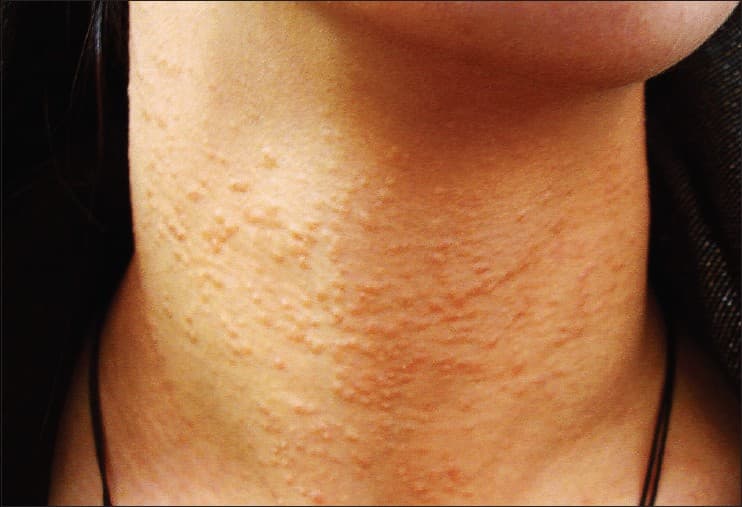
| Generalised or eruptive | Crops of many small, often brownish bumps across the neck, chest, upper abdomen, armpits and upper arms; these typically itch (DermNet, Ghanadan, Child case). | Starts younger than the localised type, often before or around puberty; more common in people with darker skin (DermNet, Ghanadan, Clear cell case). |
| Familial | Eyelid and facial syringomas that run in the family. | Appears before puberty; autosomal dominant (DermNet). |
| Associated with Down syndrome | Eyelid syringomas, sometimes with calcium deposits. | 18 to 39% of people with Down syndrome (DermNet). |
Under the microscope there is also the clear cell variant mentioned above. It is clinically indistinguishable from the common types; the difference is only visible on a biopsy, and it matters because of its link with diabetes (Ghanadan, DermNet).
Symptoms: what syringomas look like
Syringomas have a recognisable look, and a dermatologist can usually name them on sight (DermNet):
- Size: 1 to 3 mm, occasionally up to 5 mm, firm and rounded (DermNet, Ghanadan).
- Colour: skin-coloured in about half of cases, otherwise brownish, pink, yellowish or white; some look slightly see-through or cyst-like (DermNet, Yonsei).
- Pattern: solitary, or more often several in a cluster, frequently symmetrical on both sides of the face (DermNet).
- Location: the eyelids in 7 out of 10 people in one series, then the forehead (about 1 in 4), the cheeks and the genital area (about 1 in 7 each), the abdomen and the upper limbs (1 in 10 each) and the neck, 1 in 12 (Yonsei). The neck, chest and forearms are the usual sites of the eruptive type (Ghanadan). Other reported sites include the scalp, armpits and navel (DermNet).
- Sensation: usually none. About 1 in 7 people notice itching, most often brought on by sweating or summer heat, and syringomas on the vulva are more likely to itch (DermNet, Yonsei).
- Course: spontaneous regression is rare (DermNet); in one series the bumps had been present for an average of 6 years before people sought help (Ghanadan).
On brown and black skin, syringomas are often yellowish, darker than the surrounding skin or paler than it (DermNet). The eruptive form is commonly confused with mastocytosis and lichen planus (Dermoscopy cases).
The "sweat gland bumps" that people describe on the neck, chest or upper arms are usually the eruptive type. Unlike eyelid syringomas, these are easy to confuse with other conditions; in clinic series the eruptive type was usually not suspected before the biopsy (Ghanadan, Dermoscopy cases).
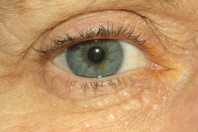
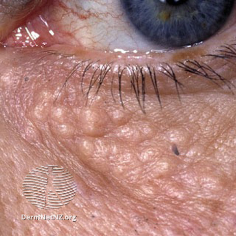
Typical syringomas: a cluster of small, skin-coloured bumps on the lower eyelid (left), and the same lesions seen close up (right).
Syringoma, milia, xanthelasma or something else?
Many small bumps can appear around the eyes and on the neck. Typical eyelid clusters are usually recognised on sight (DermNet), but the picture is less clear elsewhere: in a series of 34 biopsy-proven cases, syringoma was the first diagnosis offered in only 38% (Ghanadan), and in a series of 50 patients diagnosed clinically with syringoma, 1 in 6 turned out on biopsy to be a xanthoma (a fatty deposit such as xanthelasma) or a wart instead (Cureus 50 cases). The table lists the usual look-alikes.
| Condition | How it differs from a syringoma |
|---|---|
| Milia | Tiny pearly-white cysts of keratin sitting just under the skin surface, also common on the eyelids and cheeks. Milia are whiter and more superficial, and a dermatologist can prick their contents out through a tiny opening; syringomas are deeper, firmer and skin-coloured (DermNet milium, DermNet). |
| Xanthelasma | Soft, flat, yellow to orange patches that grow slowly over months, usually at the inner corner of the upper eyelid. Linked to blood-fat disorders, although often found with normal cholesterol (DermNet xanthoma). The most common mistaken diagnosis for the localised type (Ghanadan). |
| Plane (flat) warts | Multiple small, flat-topped, skin-coloured bumps on the face, hands and shins, caused by a virus and spread by shaving (DermNet viral wart). The most common mistaken diagnosis for the generalised type (Ghanadan). |
| Sebaceous hyperplasia | Soft yellow bumps up to 3 mm on the forehead and cheeks of middle-aged and older people, each with a tiny central dimple where the hair follicle opens (DermNet sebaceous hyperplasia). |
| Closed comedones (acne whiteheads) | Whitish follicular bumps, usually together with blackheads, papules and pustules, mostly on the face (DermNet acne). |
| Trichoepithelioma | A rare benign hair-follicle growth; a solitary lesion or multiple nodules on the face, usually inherited when multiple (DermNet trichoepithelioma). |
| Fordyce spots | Small (1 to 5 mm) yellowish or white papules on the lips and genital skin, often in crops of 50 to 100; they are visible oil glands, present in 80% of adults, and not infectious (DermNet Fordyce spots). |
| Basal cell carcinoma | A single, slowly growing, pearly or shiny nodule with fine blood vessels on its surface that may crust, bleed or ulcerate. It is a skin cancer, so a solitary growing bump near the eye should be examined rather than assumed to be a syringoma (DermNet BCC). |
| Microcystic adnexal carcinoma | A very rare sweat-gland cancer (about 5 cases per 10 million people a year) that forms a single, poorly defined, skin-coloured or yellow plaque on the central face. It has features in common with syringoma under the microscope but invades deeper, so a full-thickness biopsy is taken when a lesion is unusual (DermNet, DermNet MAC). |
| Fox-Fordyce disease | Intensely itchy bumps around the hair follicles of the armpits, pubic area and nipples, caused by trapped apocrine sweat; it occupies the same sites as some eruptive syringomas and is on DermNet's list of look-alikes (DermNet Fox-Fordyce, DermNet). |
Eruptive syringomas have also been biopsied under the assumption that they were lichen planus, granuloma annulare, sarcoidosis, histiocytosis or mastocytosis (Ghanadan, Dermoscopy cases); lichen nitidus is on the published differential list as well (Child case).
When to see a dermatologist
A confirmed syringoma is benign and never an emergency (DermNet). Having the bumps looked at serves two purposes: ruling out something else, and deciding whether removal is worth it.
- A single bump near the eye or nose that keeps growing, feels pearly, bleeds or crusts. This pattern needs examination to exclude basal cell carcinoma (DermNet BCC).
- Bumps that itch persistently, especially in the armpits, groin or on the vulva, where Fox-Fordyce disease occurs (DermNet, DermNet Fox-Fordyce).
- A spreading crop of bumps on the neck, chest or arms, which may be eruptive syringomas or one of the look-alikes above; for lesions away from the face a biopsy is recommended (Yonsei).
- You want them removed and need to know which method suits your skin tone and how likely they are to come back.
In iDerma's online consultations, typical eyelid syringomas can usually be recognised from sharp close-up photographs, and the options can be talked through from there. An online consultation with an iDerma dermatologist is a sensible first step; if a biopsy or a procedure is needed, you will be told what to arrange in person.
How syringomas are diagnosed
A dermatologist usually diagnoses eyelid syringomas by their appearance alone (DermNet). A dermatoscope, a hand-held magnifier with its own light, helps separate them from pigmented look-alikes (DermNet dermoscopy); in two reported eruptive cases it showed reticular light-brown lines, structureless light-brown areas and reticular vessels (Dermoscopy cases).
When a bump is in an unusual place, is solitary, or does not look typical, a skin biopsy settles the question. A 3 to 4 mm punch of skin is taken under local anaesthetic in a few minutes, leaving a small wound (DermNet biopsy). Under the microscope syringomas show many tiny ducts in the dermis, each with a comma-shaped or "tadpole" tail of cells, set in a firm fibrous background (DermNet, Ghanadan). Because the clinical diagnosis matched the biopsy in only about 7 out of 10 cases in one series, its authors recommend a biopsy especially for lesions away from the face (Yonsei). The sample needs to include the full thickness of the skin so that microcystic adnexal carcinoma, which can mimic syringoma at the surface but invades deeper, is not missed (DermNet).
If the pathologist reports the clear cell variant, your doctor will arrange a blood glucose test, since this pattern is linked to diabetes (DermNet, Clear cell case). No blood test diagnoses a syringoma; this glucose check is the only one the guidance recommends (DermNet).
Treatment: can syringomas be removed?
Yes, with two caveats. First, no cream, tablet or procedure makes syringomas disappear reliably; treatment aims to make them less visible with as little scarring as possible, and DermNet describes the results as often suboptimal (DermNet). Second, they tend to come back after superficial treatment, because the growth extends into the deeper skin and only the top part is removed (DermNet). Treatment is purely cosmetic, so leaving syringomas alone is a reasonable choice (DermNet, Child case).
| Method | What it involves | Points to weigh |
|---|---|---|
| Ablative laser surgery (carbon dioxide or erbium) | A focused infrared beam vaporises the top of each bump; fractional settings create microscopic areas of treatment and have largely replaced whole-surface treatment because they cause fewer complications (DermNet CO2 laser). | Listed by DermNet and the case literature (DermNet, Child case). DermNet gives about two weeks' recovery for carbon dioxide laser resurfacing in general (DermNet CO2 laser). In one study, 7 of 11 patients improved after carbon dioxide laser and 4 saw the bumps return (Yonsei). Erbium lasers heal faster with fewer side effects but do not reach as deep (DermNet CO2 laser). |
| Electrosurgery | An electrode delivers a high-frequency electric current that heats and destroys the bump (DermNet electrosurgery). | Burns are among its listed risks, and a burn can leave a small scar (DermNet electrosurgery, NHS scars). |
| Surgical excision | Individual bumps or a small cluster are cut out under local anaesthetic (DermNet). | Practical only for a few bumps or one small cluster. Every excision leaves a small scar, and pigment change after any treatment is more likely in darker skin (DermNet). |
| Cryotherapy | Liquid nitrogen freezes the bump (DermNet cryotherapy). | Listed in the case literature for eruptive syringomas (Child case). DermNet's cryotherapy page lists darker skin among the situations in which freezing should not be used (DermNet cryotherapy), and pale marks after a skin injury can be permanent (DermNet PIH). |
| Chemical peels and dermabrasion | Acid solutions or mechanical sanding remove the upper skin layers (DermNet chemical peels, DermNet dermabrasion). | Reported for widespread eruptive syringomas; results are described as often unsatisfactory (Child case). |
| Vitamin A-derived (retinoid) treatment | A prescription cream used long-term to improve the appearance (DermNet), or tablets in widespread eruptive cases (Child case). | Aims to improve the appearance rather than remove the growths; the tablets do not lower the chance of recurrence, and of four reported eruptive cases treated with them only one improved (Child case). |
| Anti-itch treatment | A prescription topical used only when the bumps itch (DermNet, Yonsei). | Calms the itch; does nothing to the bumps themselves (DermNet). |
What to expect from a procedure
Skin that has been lasered or burnt is red and crusted for a week or two (DermNet lasers, DermNet CO2 laser). A darker patch often follows and is usually temporary, although it lasts longer in darker skin; a paler patch can be permanent (DermNet PIH, DermNet lasers, DermNet CO2 laser). Raised, thickened or sunken scars and lasting pigment change are uncommon but real complications, and people who form keloid scars should say so before any procedure (DermNet, DermNet keloid, NHS scars). Asking the dermatologist to treat a few bumps first and judge how the skin heals before the whole cluster is done is a reasonable request, especially in darker skin.
Creams and home remedies
None of the guidance pages used for this article lists any over-the-counter cream, oil, acid, exfoliant or "syringoma remover" as a treatment for syringomas (DermNet). Picking, squeezing, needling or burning the bumps at home with acids or heat injures the skin without reaching the growth, and skin injury is what produces infection, post-inflammatory hyperpigmentation and small scars (DermNet PIH, DermNet lasers). If the bumps bother you, the useful question is which professional method fits your skin.
Living with syringomas and aftercare
- Sun protection: after any procedure, post-inflammatory patches darken with sun exposure, so keep the treated area out of the sun or covered with sunscreen while it heals (DermNet PIH).
- Gentle skincare: no guidance page links ordinary cleansers or moisturisers to syringomas, so there is no need to change products, but avoid scrubbing or picking the bumps (DermNet).
- Make-up and camouflage: a dedicated skin camouflage cream, long-lasting and often water-resistant, can be used to conceal marks and discolouration (BAD skin camouflage).
- Itch: the itch is linked to sweating and summer heat (DermNet, Yonsei), so staying cool and avoiding heavy sweating is the practical first step; a dermatologist can prescribe a topical for the itch if that is not enough (DermNet).
- Follow-up: if you have had syringomas removed, new bumps in the same area are not unusual, because recurrence after superficial treatment is documented (DermNet); they are just as harmless as the originals, and whether to treat them again is a cosmetic choice (DermNet).
Can syringomas be prevented?
No. There is no known way to stop syringomas from forming, because their trigger is unknown and in some families the tendency is inherited (DermNet). No guidance page or study links diet, skincare or hygiene to them, and avoiding sweating helps only the itch (DermNet, Yonsei). What you can influence is the outcome of treatment: a method suited to your skin tone, and sun protection afterwards, give the best chance that the treated skin looks better than the bumps did (DermNet, DermNet PIH).
Syringomas in children
Children do develop syringomas, including the inherited eyelid type and the eruptive type on the neck and trunk; ages from 5 upwards are reported (DermNet, Ghanadan, Child case). In children the priority is the correct diagnosis, since the eruptive form is easily mistaken for flat warts, acne or an itchy rash (Child case). Treatment is seldom needed in childhood, because the bumps are harmless and procedures can scar; in a published case the eruption of a 6-year-old improved without any treatment (Child case). A review of 42 reported eruptive cases found that most were simply observed (Child case).
If you are unsure whether the bumps on your or your child's face or neck are syringomas, an online consultation with an iDerma dermatologist is a practical first step: typical eyelid syringomas can usually be recognised from photographs, and you will be told whether a biopsy or any treatment is worth considering.